We offer resources, support, and mentorship to world-class biomedical researchers at the University of Michigan, and across the state.
Our mission at Fast Forward Medical Innovation (FFMI) is to provide the expertise and knowledge to help access and navigate the unparalleled biomedical innovation resources at the University of Michigan.
Whether you’re a student, faculty, potential collaborator, or industry partner, we encourage you to connect with a Fast Forward Medical Innovation team member to explore our program's unique offerings and find your path to groundbreaking innovation.
FFMI is part of a rich ecosystem of opportunities available to biomedical researchers at the University of Michigan. Together with our collaborators, we work to create a robust network of resources that help drive innovation to successful commercialization. The FFMI team is here to provide mentorship and guidance to the abundant funding, education, and support structure resources that will benefit your research. We look forward to working with you!
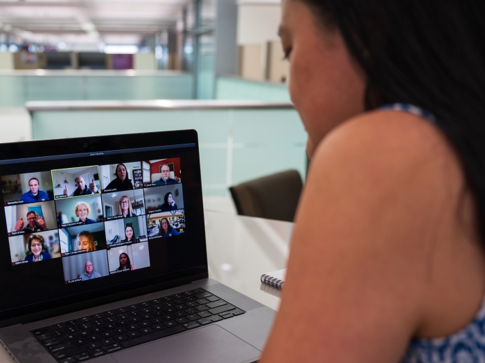
Don't miss your chance to participate in this project-based, experiential course designed to help academics launch new innovations, including medical devices/diagnostics, digital solutions, drugs, educational/training interventions, research tools, and many others.
Register by Wednesday, May 1, for the Spring 2024 Cohort, which begins with a virtual kickoff event on Friday, May 10, from 12:00 PM - 4:30 PM.
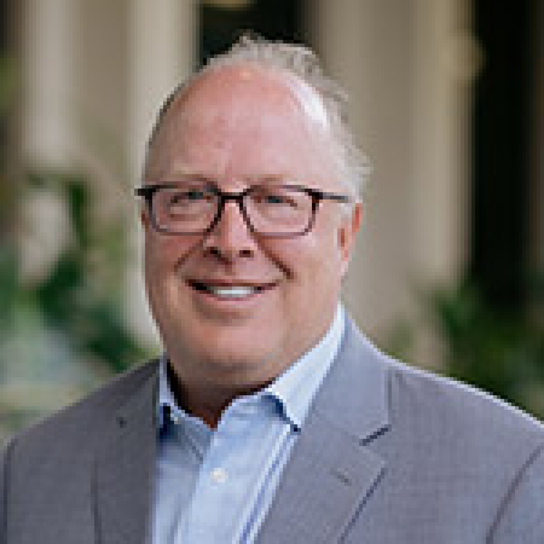
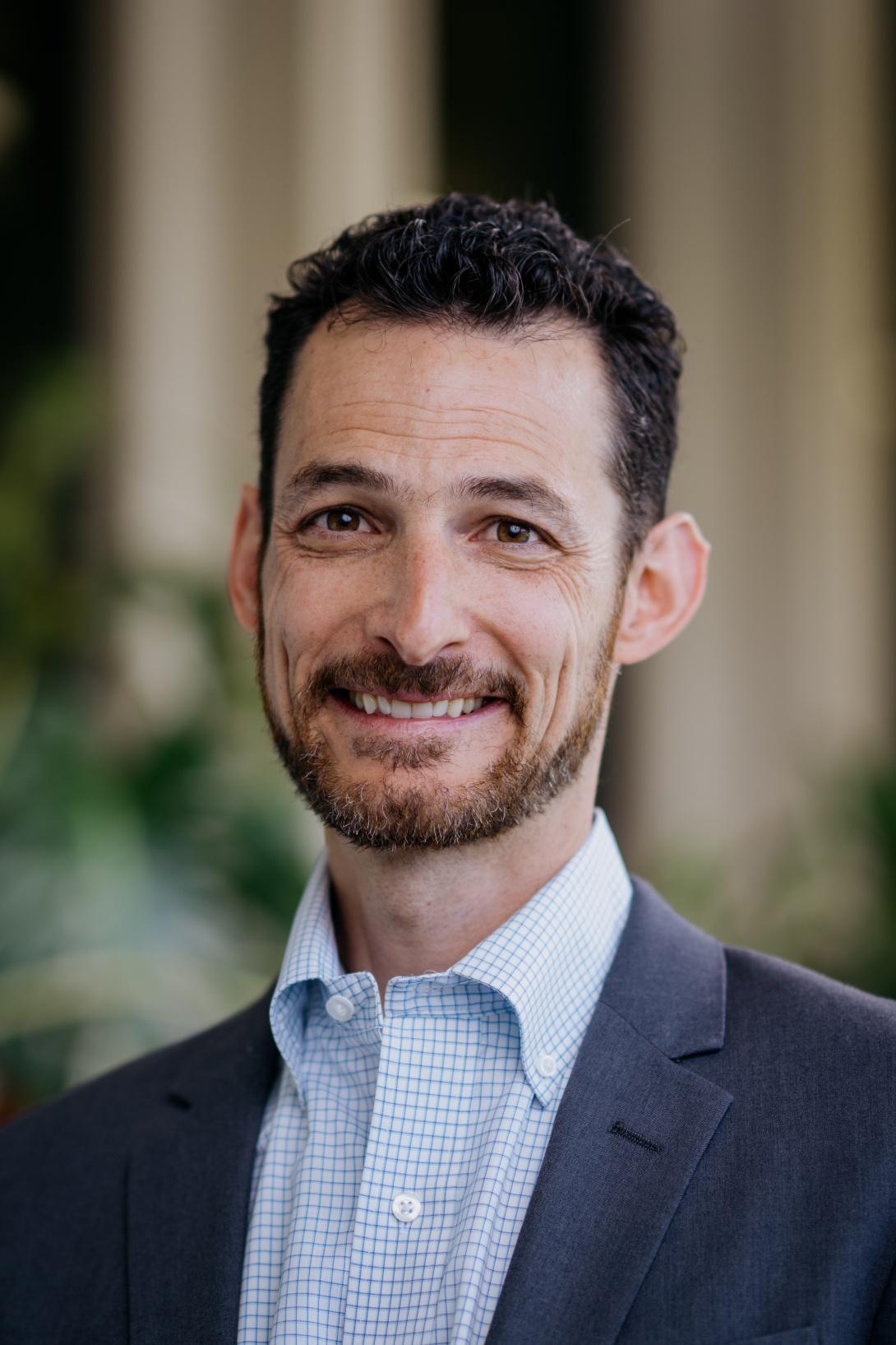
Prior to becoming Director of FFMI, Brad led the MTRAC Hub for Life Sciences. He has served as a Mentor in Residence for U-M Innovation Partnerships, was the Director of Product Development at Aastrom Biosciences, and the Director of Cardiac Research at Osiris Therapeutics Inc. He also spent several years as a Principal Scientist with Pfizer’s Esperion Division. Brad has 15-plus years of experience in the development of drugs and biologics, and particular expertise in preclinical development and translational research in the field of cardiovascular physiology and regenerative medicine.
Brad received in PhD in Physiology in 1993 from the University of Michigan, and completed post-doctoral fellowships in the departments of Cardiothoracic Surgery and Physiology at the University of Wisconsin.
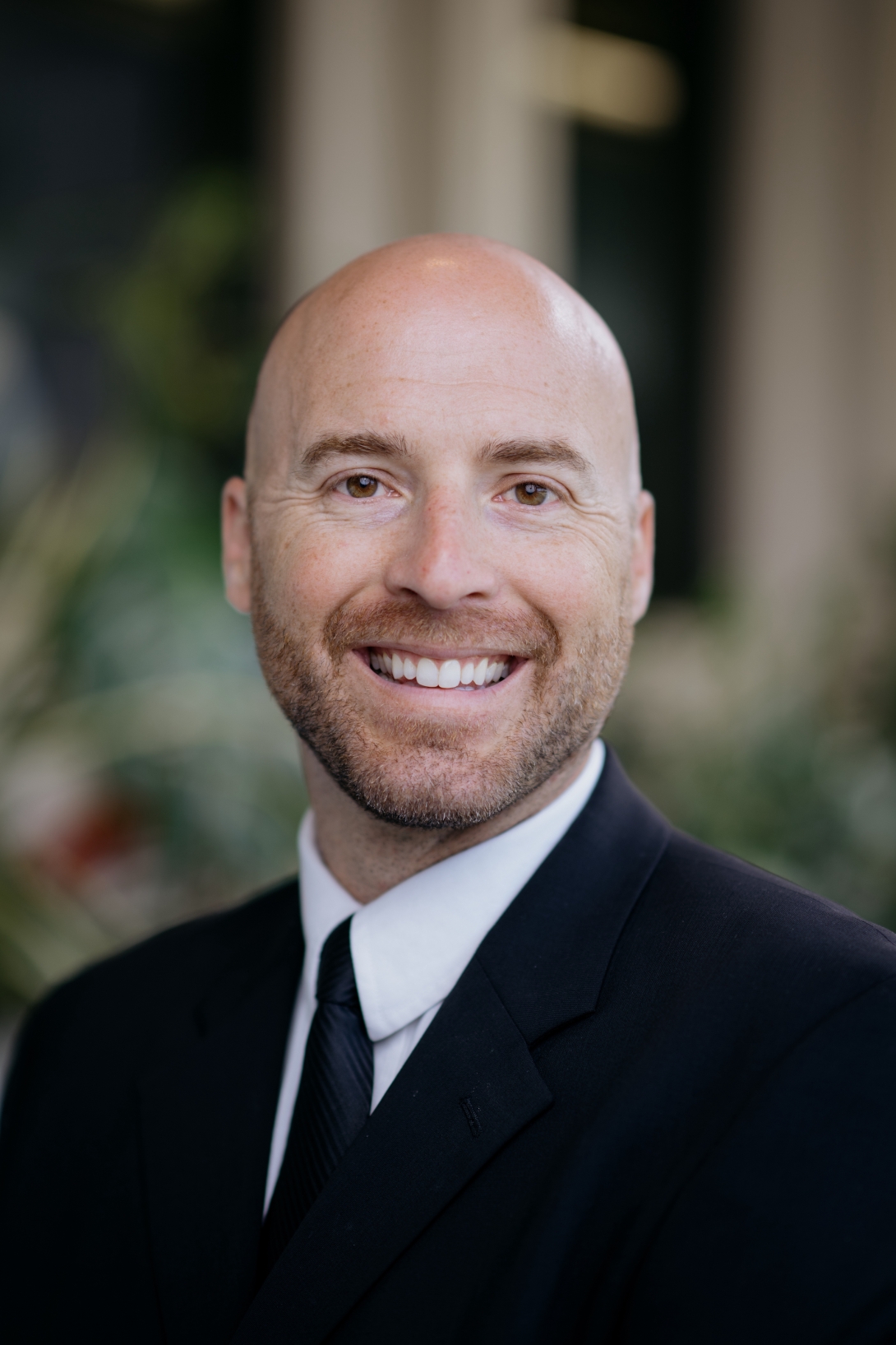
Mike leads the FFMI Business Development team. The team works with Industry partners and U-M faculty to enable and catalyze productive academic-industry partnerships, leveraging innovations to improve healthcare. He utilizes over 15 years of healthcare research experience to establish mutually beneficial deals with industry to further the mission of the healthcare system.
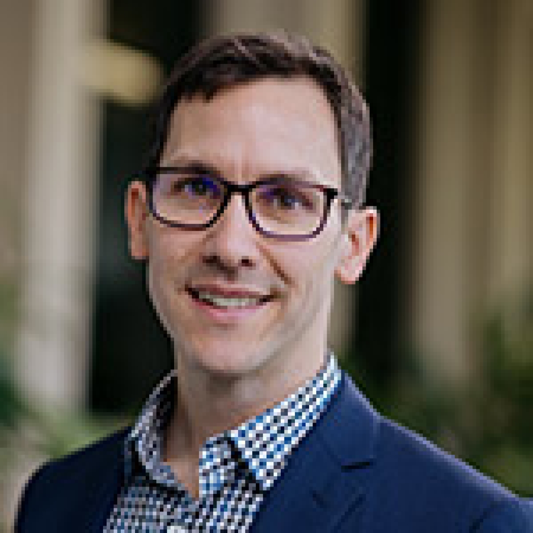
Jon joined the FFMI team in 2014 as the Director of Commercialization Education where he develops and leads biomedical innovation, entrepreneurship, and commercialization education and training programs for U-M Medical School faculty, staff, and trainees – resulting in education and development training for more than 1,000 participants and 50 emerging biomedical technologies per year.
He has a doctorate in education from the University of Michigan-Dearborn, as well as a master’s in education from East Carolina University and a bachelor’s degree from Adrian College.
John joins U-M after 17 years in the med tech industry. Most recently, he was the Director of Clinical Engineering at a breast cancer screening startup (Delphinus), leading the development of medical image review and analysis software. John started his career at GE Healthcare, with roles in advanced applications, clinical research, molecular imaging, and led the development of features for non-invasive coronary artery CT imaging.
John worked as a Venture Associate evaluating startups for the Zell-Lurie Fund, and is pursuing a part-time MBA at U-M/Ross. HE holds a BSE in Electrical Engineering and MSE in Biomedical Engineering.
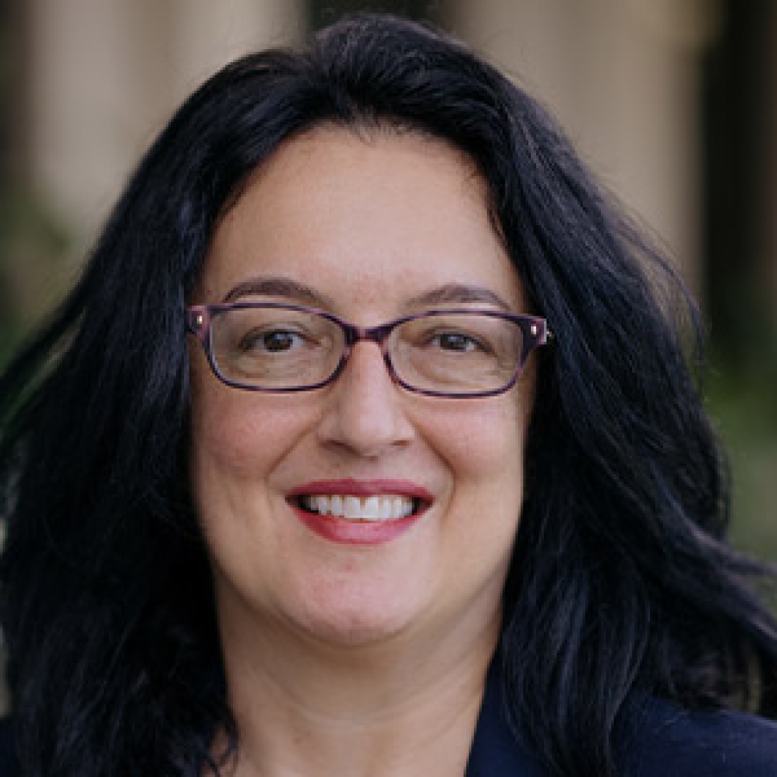
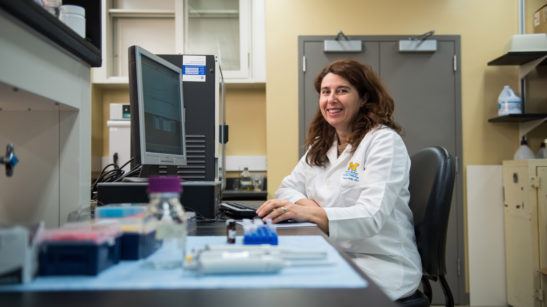
Prior to joining FFMI, Emilija worked for the U-M Rogel Cancer Center since 2006. She brings over 10 years of experience working with oncology clinical trials.
Emilija received her M.D. from the Medical School at the University St. Cyril and Methodius in Macedonia.
Prior to joining FFMI, Shelby worked with both FFMI and the Cancer Center through her participation in the Research Administration Fellowship Program based in the Medical School Office of Research.
Shelby completed her Ph.D. and Postdoctoral research training in cancer research at the University of Michigan.

Prior to joining FFMI, Ingrid worked at the Hospital for Sick Children in industry partnerships and commercialization, and the University of Windsor’s Office of Research and Innovation overseeing the health research portfolio.
Ingrid completed her Ph.D. in molecular biology and M.B.A. at the University of Windsor.
Ashley Schork joins FFMI after spending over six years working at the University of Michigan Life Sciences Institute (LSI) administratively managing several research centers in the areas of structural biology, cryo-electron microscopy, drug discovery, and high-throughput screening. Prior to the position at LSI, Ashley worked at Arbor Research Collaborative for Health providing administrative support to NIH and PCORI funded nephrology studies.
Ashley has both her BA and a Masters in Non-Profit Management from DePaul University.
Katherine joins FFMI from the Center for Academic Innovation (CAI), where she served as a Learning Experience Design Student Resident and Online Student Services Fellow during her graduate program. Prior to her work at CAI, she worked at U-M’s Office of Undergraduate Admissions where she was involved in developing training opportunities and materials.
Katherine holds a bachelors, masters, and graduate certificate from the University of Michigan.
Charlotte comes to FFMI from Physical Medicine & Rehabilitation MedRehab, where she was an administrative assistant for the ACU manager.
Charlotte holds a master’s in health administration from Southern New Hampshire University, and an undergraduate degree from Michigan State University.
Associate Professor, Obstetrics and Gynecology
Project: Novel cell therapy targets premature ovarian insufficiency
Medical Director, Neurosurgery
Professor, Otorhinolaryngology
Project: Device to Help Stroke Patients
Medical Director, Pediatrics-Cardiology
Section Head, Pediatrics-Cardiology
Project: Nerve-Controlled Prosthesis
Section Head, Obstetrics and Gynecology
Project: Device to Predict Preterm and Term Birth
Project: Prediction of Cancer Recurrences with Biomarkers
Project: Novel human developmental toxicity testing platform

Professor, Microbiology and Immunology
Project: C3d Immunotherapies: A Novel Platform to Treat Cancer
Professor, Chemical Engineering
Professor, Macromolecular Science and Engineering
Project: Novel targeted treatment for lung injury
Associate Director of Academic Programs - Molecular and Physiology
Research Professor - Internal Medicine - Institute of Gerontology
Project: Seq-Scope - Providing finer spatial resolutions for basic and clinical researchers
Project: Novel treatment for osteoarthritis
Project: Michigan Tinnitus Device
Service Chief, Surgery, Plastic Surgery
Section Head, Surgery, Plastic Surgery
Project: Nerve-Controlled Prosthesis
Professor, Internal Medicine - Cardiology
Project: Novel Strategy Offers Improved Treatment for Arterial Plaque Calcifications
Project: EVOQ Therapeutics- Delivering immunotherapies to improve the lives of individuals fighting autoimmune disease
Get a monthly digest of medical research and innovation, straight to your inbox.
Building 520, 3rd Floor
2800 Plymouth Road
Ann Arbor, MI 48109-2800